Impacted Wisdom Teeth: How Nerve and Sinus Proximity Are Assessed
A patient-friendly explanation of how anatomy, imaging, and surgical options are discussed when wisdom teeth appear close to a nerve or sinus.
Quick answer: When an impacted lower wisdom tooth appears close to a nerve, or an upper wisdom tooth is near the maxillary sinus, the clinician combines symptoms, examination, and appropriate imaging to assess options and explain risk. “Close” on an image does not by itself predict an outcome or require one specific procedure.
These anatomical questions often worry patients who are planning care abroad. A useful consultation should translate the image into an individual discussion: what structure is nearby, what uncertainty remains, what alternatives exist, and what follow-up is required.
The lower-jaw nerve question
The inferior alveolar nerve travels through the lower jaw and supplies sensation to areas including the lower lip and chin. Roots of a lower wisdom tooth can appear near its canal on dental imaging. Ask the clinician to show you the relationship and explain whether additional information would change the plan.
Temporary or persistent altered sensation is one of the risks that may be discussed for lower wisdom tooth surgery. Your individual risk cannot be calculated from a generic article. It depends on the anatomy and proposed procedure, and the surgeon should explain it in understandable terms.
The upper-tooth and sinus question
Upper wisdom teeth may be near the floor of the maxillary sinus. Imaging may help the clinician assess that relationship along with the tooth’s position and surrounding structures. Ask whether sinus proximity changes the procedure, recovery instructions, or warning signs relevant to your case.
Tell the team about current sinus symptoms, previous sinus surgery, and recent respiratory illness. That information does not replace examination, but it may matter to scheduling and planning.
When three-dimensional imaging may be considered
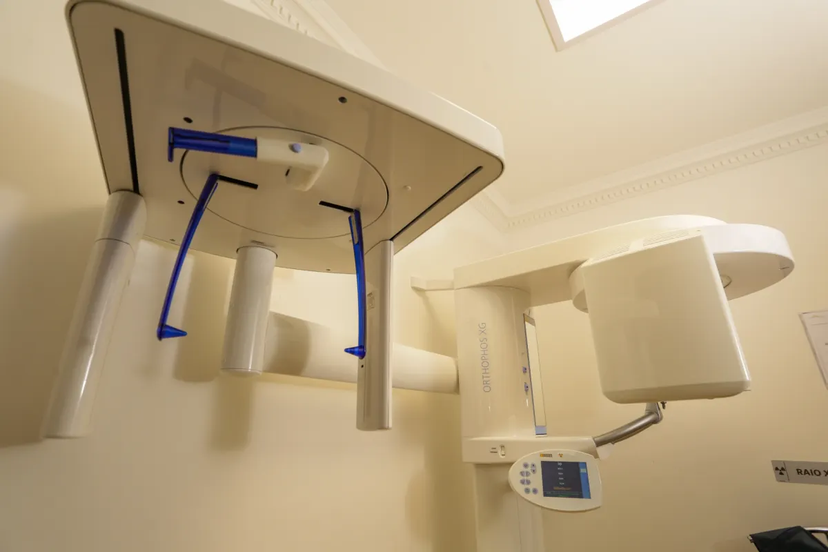
The FDA explains that dental CBCT can provide three-dimensional information for selected clinical questions and should be performed only when necessary. Ask what a CBCT would clarify beyond existing images and whether it could change the options.
Keep copies of previous imaging and dates. Providing original files rather than screenshots may help the team judge whether they are usable and avoid unnecessary repetition.
Ask about alternatives and the decision pathway
Depending on the clinical situation, the discussion may include removal, monitoring, or another technique. For selected lower wisdom teeth, a surgeon may discuss coronectomy, in which the crown is removed while roots are intentionally retained. It has its own indications, risks, and follow-up; it is not a universal safer substitute.
The 2024 AAOMS clinical paper emphasizes evaluation of disease, anatomy, risks, and monitoring when an impacted tooth is retained. Ask what evidence supports the proposed option and what future review would be needed.
Plan travel around uncertainty and review
Do not use the image alone to choose a procedure date or return flight. Confirm the evaluation, anesthesia plan, expected early review, and what could delay travel. Arrange an escort if sedation is used and follow individual instructions about activity and transport.
Before leaving São Paulo, request a procedure summary, copies of relevant images, written aftercare, and instructions for altered sensation, sinus-related concerns, bleeding, swelling, fever, or increasing pain. Ask which concerns require urgent local care.
For the broader process, read Wisdom Tooth Removal in São Paulo and the international patient FAQ, or contact Hori Clinic about an evaluation.
Compare observation and removal as complete plans
When removal is recommended, ask what specific finding supports it, what could happen with observation, and how each option would be monitored. A tooth that is not removed may still need periodic clinical or radiographic review. The interval should come from the treating clinician and your individual findings, not a generic online schedule.
If surgery is proposed, ask how the relationship to the nerve or sinus affects the technique, consent discussion, recovery, and follow-up. Clarify which symptoms are expected and which require prompt assessment. International patients should leave with copies of the relevant imaging and a written procedure summary so that a professional at home can understand the site, the decision, and any concern that appears after travel.
Educational information only. This article is not a diagnosis or personalized treatment recommendation and does not replace an evaluation by a qualified professional.
Related Articles
- Digital Planning for Jaw Surgery and Dental Implants: What the Technology Adds
- Orthognathic Surgery in Brazil: Planning and Recovery for International Patients
- TMJ Pain and Difficulty Opening the Mouth: What an Evaluation May Include
- Wisdom Tooth Removal in São Paulo: A Guide for International Patients