Digital Planning for Jaw Surgery and Dental Implants: What the Technology Adds
CBCT, intraoral scans, and virtual planning can improve communication and preparation, but they remain tools within an individual clinical decision.
Quick answer: Digital planning combines records such as photographs, intraoral scans, X-rays, or CBCT with software used to study anatomy and prepare treatment. These tools may improve visualization, communication, and coordination, but they do not replace diagnosis, informed consent, surgical judgment, or follow-up.
International patients often send digital records before travel. That can make an appointment more organized, but it is important to understand what each file shows and what still requires confirmation in person.
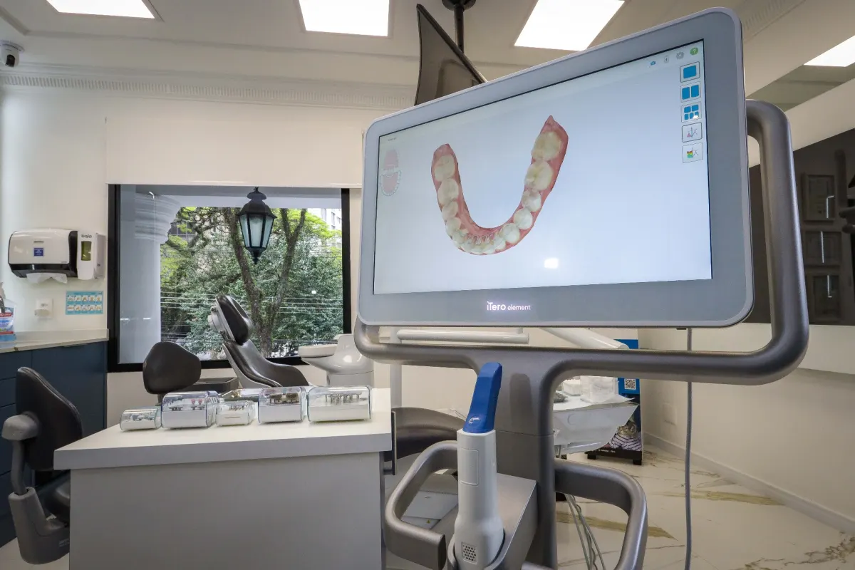
Intraoral scans create a digital surface model
An intraoral scanner records the visible shape of teeth and oral structures. The resulting model may support restorative design, bite records, orthodontic planning, and communication with a laboratory. Ask whether an existing scan can be imported and how recent it needs to be.
Because it records surfaces, an intraoral scan does not provide the same information as an X-ray or CBCT about bone and internal anatomy. One technology does not make every other record unnecessary.
CBCT provides selected three-dimensional information
Dental cone-beam computed tomography rotates around the patient and reconstructs a three-dimensional image of dental and maxillofacial structures. The FDA’s CBCT guidance says it can support diagnosis and planning for selected conditions, including implant planning, abnormal teeth, jaws, face, and trauma.
CBCT uses ionizing radiation. It should be justified by a clinical question and optimized to obtain adequate information while limiting unnecessary exposure. Ask why it is recommended, whether a lower-radiation or existing exam can answer the question, and how the result may change care.
Virtual planning organizes clinical decisions
Software may combine dental models and imaging to evaluate implant position, jaw movement, bite relationships, or the design of guides and splints. The team may use the plan to coordinate with orthodontists, restorative dentists, laboratories, or hospital staff.
Ask who reviews and approves the virtual plan and how accuracy is checked. A colorful simulation is not proof of candidacy or a guaranteed preview of healing and appearance.
Guides and printed devices have defined jobs
For selected procedures, a digital plan may be transferred to a surgical guide, splint, model, or temporary restoration. Ask what device is being made, what it controls, what it does not control, and what the clinician will do if the operative findings differ from the plan.
Technology should support a safe decision pathway rather than lock the team into a plan that no longer fits the clinical situation.
Share files securely and keep your own copy
Ask which file formats the clinic needs and use a communication channel confirmed by the clinic. Avoid sending sensitive health records to an unverified social media account. Keep original files, reports, and dates rather than only screenshots.
After care, request the records needed for follow-up at home. For implants, the FDA advises patients to keep information about the implant system. For jaw surgery, ask for operative and discharge summaries, relevant imaging, and any plate or device details the team considers appropriate.
Use technology to ask better questions
During the consultation, ask the clinician to show the problem being evaluated, the options considered, the proposed position or movement, important nearby anatomy, uncertainties, and how the plan connects to the final restoration or bite. Then discuss risks, alternatives, travel timing, and follow-up in language you understand.
For related reading, see dental implant planning, orthognathic surgery in Brazil, and the international patient FAQ. To ask about an evaluation at Hori Clinic in São Paulo, contact the team.
Ask to see how the digital record changes the plan
Technology is most useful when it answers a defined question. During the consultation, ask the clinician to point out the relevant anatomy, proposed position or movement, margin of uncertainty, and the choices that were considered. Ask whether the same decision could be made with existing records and whether a new scan would expose you to radiation or add cost.
For international care, request copies of the original image files and reports, not only screenshots from planning software. Confirm who owns or stores the virtual plan, how long it is retained, and what can be shared with your professional at home. If a guide, splint, or temporary restoration is manufactured, ask how the team checks its fit and what happens if current clinical findings differ from the digital model.
Educational information only. This article is not a diagnosis or personalized treatment recommendation and does not replace an evaluation by a qualified professional.